Microscopic engines driving life
This article explores the world of molecular motors, particularly highlighting the contributions of Ronald Vale, Investigator, HHMI's Janelia Research Campus, and the 2023 speaker of the TNQ Distinguished Lectures.
Rohini Karandikar
Consultant,
TNQ Foundation
12-August-2025
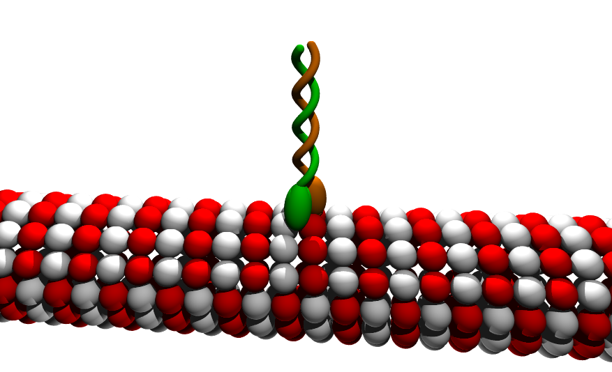
Cartoon of a kinesin dimer. Source: Wikimedia commons. CC-BY-SA 3.0 Link:https://commons.wikimedia.org/wiki/File:Kinesin_cartoon.png
Movement is an essential function in all forms of life. From microscopic organisms to a gigantic whale, any movement of or within an organism is driven by proteins called ‘molecular motors.’ These motors transport molecules, also known as cargo, along tracks or ‘highways,’ exactly the way vehicles transport people or parcels within a city. The motors also enable movements of ciliated and flagellated cells or organisms. Some motor proteins also cause filaments to slide against each other, producing a force that leads to processes such as muscle contraction and cell division.
Myosin, kinesin, and dynein are the three molecular motors that enable cellular movements. These proteins are fueled by energy from ATP hydrolysis, thereby converting chemical energy into mechanical energy that drives movement.
Myosin
Myosin is the first molecular motor to be discovered. It plays a key role in muscle contraction. Skeletal and cardiac muscles are made of contractile units called sarcomeres. Under the electron microscope, each sarcomere shows distinct and alternating light and dark bands. The light bands are composed solely of actin, while the dark bands comprise both actin and myosin. During muscle contraction, myosin interacts with actin, another protein, and also binds ATP, hydrolysing it. The ATP hydrolysis drives the sliding of actin filaments, leading to muscle contraction.
Due to its vital role in muscle contraction, including the cardiac muscles, drugs targeting myosin show a promising effect in treating heart and neuromuscular diseases. Drugs called Myotropes are myosin activators. They improve the actin-myosin interaction, leading to enhanced sarcomere contraction in patients with heart disease. One example of a Myotrope is Omecamtiv mercabil, a drug developed by Cytokinetics in collaboration with Amgen.
Ronald Vale, Investigator at Howard Hughes Medical Institute’s (HHMI) Janelia Research Campus, is also a co-founder of Cytokinetics and was the 2023 speaker of the TNQ Distinguished Lectures, where he presented a talk on Marvelous Molecular Motors.
While Omecamtiv mercabil is a myosin activator, Mavacamten—a myosin inhibitor— is used to treat a condition called hypertrophic cardiomyopathy, in which the heart muscle abnormally thickens, making it harder for the heart to pump. This disease is observed among athletes and can cause sudden death. Mavacamten blocks myosin ATPase, preventing excessive actin-myosin crossbridge formation, and eventually improving cardiac function. Mavacamten was initially developed by Myokardia Inc. (later acquired by Bristol Myers Squibb).
Kinesin
While myosin moves along actin filaments, kinesin is a motor protein that typically moves along different tracks—microtubules—transporting cargo from the nucleus towards the cell surface, or the ‘plus end’. Kinesin was discovered by Vale and his colleagues in 1985 from the giant axon of the squid. Deriving energy from ATP hydrolysis, kinesin performs a ‘hand-over-hand’ movement as it walks along microtubules.
Vale’s team developed an in vitro mobility assay for direct visualisation of fluorescently labelled kinesin molecules moving along a microtubule. The assay has been in use for nearly three decades now.
Kinesins play a major role in mitosis, positioning of membrane-enclosed organelles, transport of vesicles, development and function of neurons, axonal transport, and microtubule dynamics, among other functions.
Dynein
Dynein is a motor protein discovered in 1965 by Ian R Gibbons, 20 years before kinesin was discovered. Unlike kinesin, dynein transports cargo to the cell center, also known as the ‘minus end’. Dyneins are broadly classified into two types: axonemal and cytoplasmic. Axonemal dyneins drive the motility of cilia and flagella, while cytoplasmic dyneins transport cargo towards the minus end of a cell, and are particularly important in transporting material from the axon terminal to the neuron’s cell body.
Dynein has a more complex structure compared to kinesin, with a molecular weight of ~1.4 MDa. For a long time, its large size presented challenges in its expression, purification, and manipulation. However, advances in imaging, such as cryo-electron microscopy (cryo EM) have made it possible to unravel the mechanism by which dynein enables movement.
Gibbons’ discovery set the stage for further investigation of dynein. Building on the discovery, Vale’s group was the first to express and purify yeast cytoplasmic dynein. Vale’s lab also determined the structure of dynein’s microtubule-binding domain and motor domains. Their work has paved the way for investigating dynein’s movement, regulation, and neurodegenerative diseases such as Alzheimer’s and Parkinson’s that can result from dysfunctional dynein.
Molecular motors belong to a superfamily of motor proteins that orchestrate movements inside a cell. Research on their function and mechanisms has revolutionised our understanding of intracellular transport and the role of molecular motors in diseases. It has also enabled the design of targeted therapies for a wide range of diseases.